Home / Research / Core Facilities /
 The Brain Imaging and Electrophysiology Core (BIEC) helps researchers studying intellectual and developmental disabilities (IDD) by providing technical and scientific support for brain imaging. They offer access to advanced imaging methods like MRI and electrophysiology.
The BIEC works closely with other research units and centers at the UW Department of Radiology and the UW Center for Human Neuroscience.
Core Services:
- Expert guidance in designing brain imaging studies and selecting appropriate methods.
- Training and assistance in preparing research subjects, especially young children with IDD.
- Developing specialized equipment and imaging protocols for specific studies.
- Training in the safe use of imaging equipment and techniques.
- IT support for managing images.
- Training and assistance in data collection and advanced image analysis.
- Help with preparing Human Subjects IRB applications for neuroimaging research.
The BIEC is divided into three components:
- Technical MRI and experiment development.
- Image post-processing and analysis.
- Human electrophysiology.
Each component is led by an expert who collaborates closely to support researchers effectively.
Brain Imaging and Electrophysiology Core

Brain Imaging and Electrophysiology Core Breakdown
The overall goal of the Core is to support affiliates and train junior investigators to perform scientifically sound research by providing a variety of (and selecting appropriate) advanced imaging modalities such as structural and functional/physiologic MRI, multinuclear MR spectroscopy, ERP/EEG, MEG, and PET as well as by designing study-specific sophisticated analysis tools for both human and animal studies.
The overall goal of the Core is to support affiliates and train junior investigators to perform scientifically sound research by providing a variety of (and selecting appropriate) advanced imaging modalities such as structural and functional/physiologic MRI, multinuclear MR spectroscopy, ERP/EEG, and MEG as well as by designing study-specific sophisticated analysis tools for both human and animal studies.
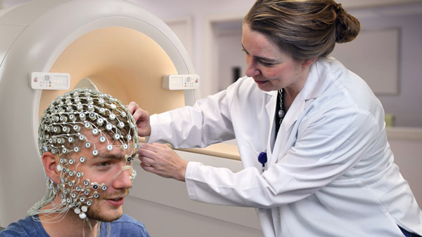
A central resource of the Human Electrophysiology Component (HEC) is the Electrophysiology Laboratory, also housed in core space providing tight integration of HEC imaging services with MR image acquisition. This facilitates easy collaboration between these two brain imaging modalities and is especially important in support of the laboratory’s in-magnet EEG system. This system, consisting of a BrainAmp MRplus MRI-compatible 64-channel digital system (Brain Products, Inc.), permits simultaneous EEG and fMRI data acquisition. Use of this system in the magnet is complex and requires specialized support from HEC staff to help prepare the subject as well as to monitor acquisition to ensure a good EEG data set during the imaging experiment. IDDRC affiliates also have access to an EEG/event-related potentials (ERP) apparatus (Electrical Geodesics Inc., Eugene, OR) that utilizes the dense array electrode system that records brain activity from a 128 scalp electrode array.
The lab currently has ten 128 channel nets of various adult, child, and infant sizes. The laboratory can also record a wide range of autonomic measures in conjunction with EEG for use in studies of attention, stress, and emotion.