Home / Stories /
 by Kate Forster
February 7, 2017
The participants come to McLaughlin’s lab for a total of three visits. On the first visit, they are interviewed, and they fill out questionnaires designed to assess their environmental experiences, family relationships, and the degree of violence they experienced in different areas of their lives. They also perform a variety of behavioral tasks designed to gauge how they learn about and respond to threat and safety cues.
The second session focuses on an assessment of mental health. This includes a number of structured clinical interviews to determine how disruptions to some of the emotional processing systems in the brain might increase the risk for mental health problems like anxiety, depression, and PTSD in children who have experienced trauma.
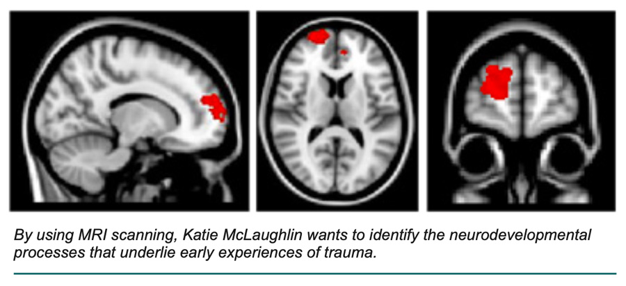
The third session involves functional magnetic resonance imaging (fMRI), where McLaughlin observes which regions of the brain become activated as participants perform a variety of emotion processing tasks. In one task, participants look at faces with different types of emotions while McLaughlin measures how different areas of the brain respond to facial cues. “By looking at how the participants respond to faces that seem angry or scared, which could suggest a threat, we’re measuring basic reactivity to different emotional stimuli,” she said. “Facial cues of emotions are very important signals that there is something threatening in the environment and that it should be paid attention to.” By understanding how regions of the brain that detect and learn about threats respond to these cues, McLaughlin hopes to gain insight into how these brain regions may develop differently in children who have experienced significant environmental threats.
Another task involves learning to associate something neutral, like a triangle, with something threatening, like a loud noise. For this task, McLaughlin wants to observe how systems in the brain develop and extinguish fear responses to cues that have previously been associated with threat. “We’re specifically interested in the amygdala—the region of the brain that’s involved in identifying salient cues in the environment and helps us learn about threats,” she said. Examining the amygdala during this kind of learning task will provide information about whether children who have been exposed to violence learn about new potential threats more quickly or have a more difficult time extinguishing fear responses to cues that were previously associated with threat but are now safe, compared with children who have never experienced violence. “We expect that participants who have been exposed to trauma will retain that fear memory for a longer period of time,” she said. If this is the case, it would suggest new avenues for preventing the onset of anxiety and PTSD in children who have experienced violence.
McLaughlin is looking not only at these types of automatic emotional regulation processes but also at effortful forms of emotion regulation. An example of this is something called cognitive reappraisal, which involves thinking about a situation in a different way to change its emotional significance. In another task, children view images that are negative. In some trials, they simply view the images without trying to change their emotional response. In other trials, they use cognitive reappraisal strategies learned before the scan to try to decrease their emotional response by thinking about the picture in a different way. If differences emerge between children with and without exposure to violence, it could provide important information for intervention, as cognitive reappraisal is a tool frequently used in treatment of children who have experienced trauma.
 McLaughlin is also gathering information about the structure of the brain, including the size of different regions, like the amygdala, the hippocampus, and different parts of the prefrontal cortex, and she is looking at white matter tracts, which connect one part of the brain to another and form the connections between different nodes and networks of the brain. She is also examining functional connectivity between different brain regions when participants are not performing any tasks at all. “Even when you’re resting, you should see stronger relationships between the patterns of activity in particular regions. It’s like a signature that stays with you whenever those regions have been activated together a lot in the past. By looking at what’s correlating with what, even when you’re resting, we can get a sense of how different

